

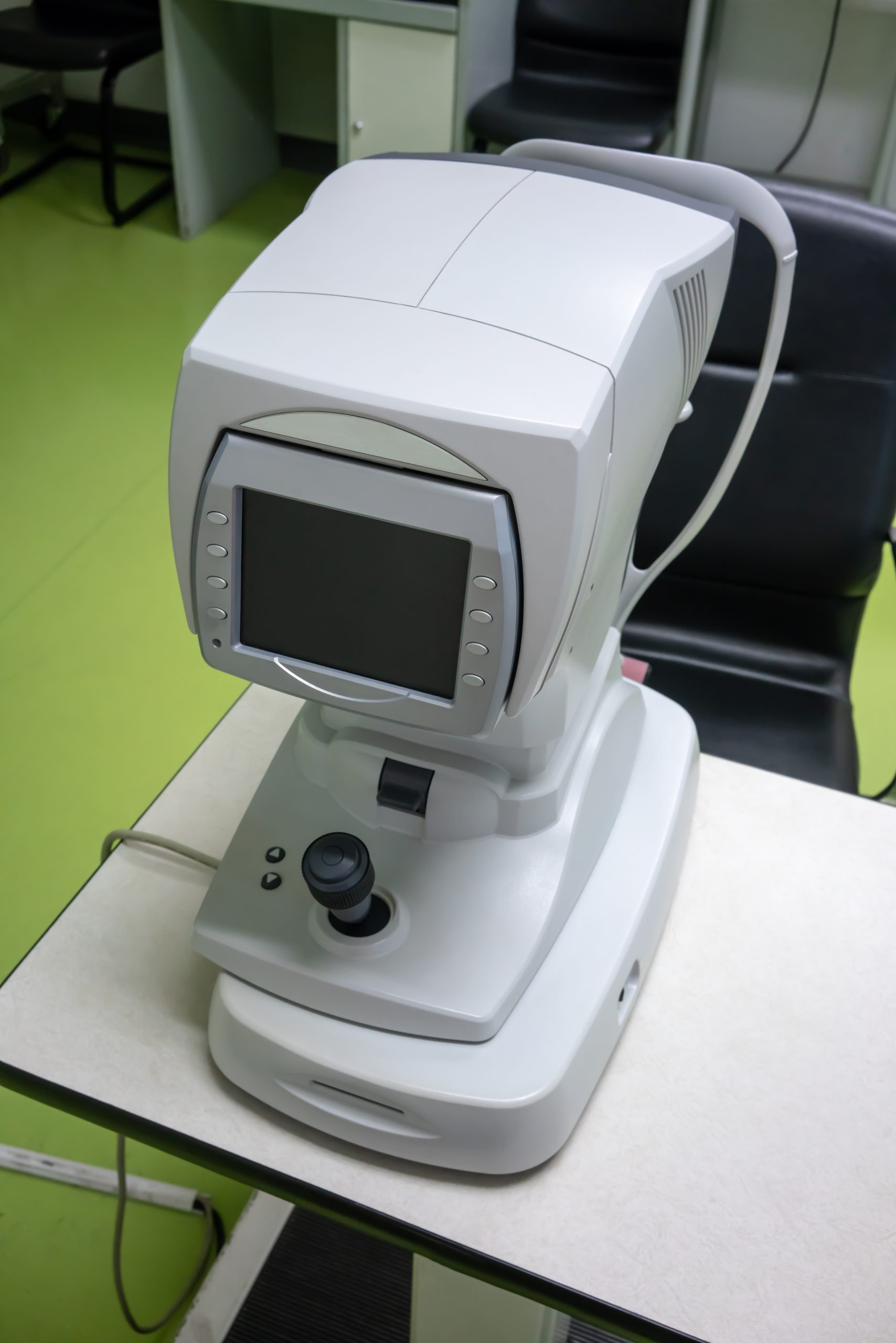
Corneal topography is one of the more fascinating pieces of diagnostic technology in modern eye care — it creates a detailed, color-coded map of the front surface of your eye, revealing details a standard exam can't catch on its own.
What Is the Cornea, and Why Map It?
The cornea is the clear, dome-shaped front surface of the eye, responsible for most of its focusing power. Even small irregularities in its shape can significantly affect vision, and some conditions specifically alter corneal shape over time.
How Corneal Topography Works
A topographer projects a pattern of light onto the cornea and analyzes how it reflects back, generating a detailed elevation map — similar in concept to a topographic map of terrain. The result shows curvature, thickness variations, and irregularities across the entire corneal surface.
What It's Used For
- Detecting keratoconus, a progressive thinning and bulging of the cornea, often before it's noticeable on a standard exam
- Planning LASIK, PRK, and other refractive surgery, ensuring the cornea is a good candidate and mapping the precise correction needed
- Fitting specialty and scleral contact lenses for irregular corneas
- Monitoring corneal changes over time in patients with certain conditions
- Evaluating astigmatism with more precision than manual measurements alone
What to Expect
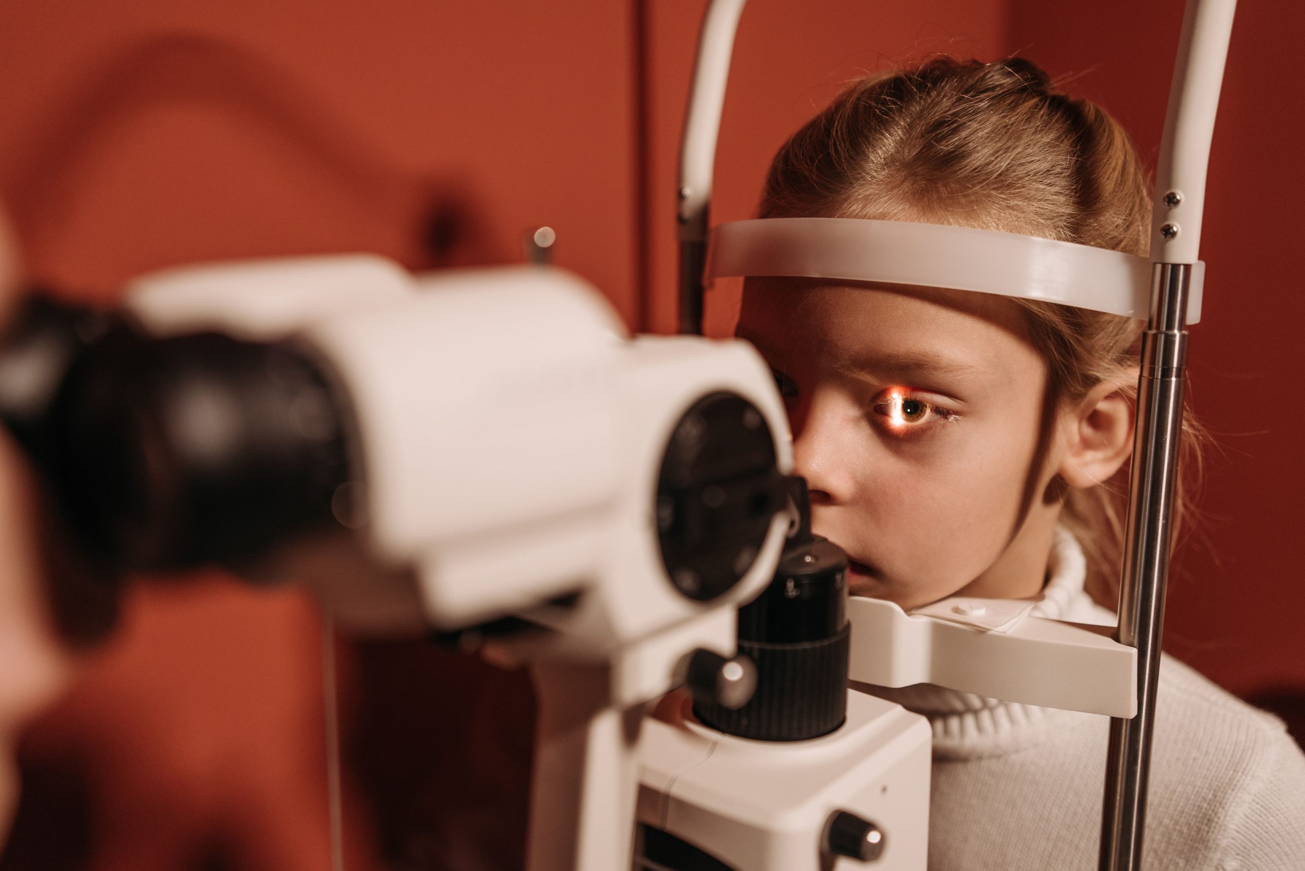
The test is quick, painless, and non-invasive — you simply look into the device for a few seconds while it captures the corneal surface. No dilation or numbing drops are required.
The Bottom Line
Corneal topography lets your doctor see subtle details about your eye's shape that directly affect both your vision and your candidacy for certain procedures.
Curious about corneal topography or considering refractive surgery? Schedule a consultation with our team.
0 Comments